Features
Summary
Findings
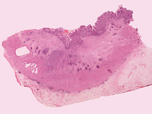
This image shows a portion of the small intestine that is markedly thickened due to inflammation, edema and fibrosis. Note that foci of inflammation is distributed in all the layers (transmural in distribution). A small fissure is beginning to extend from the mucosa to the submucosa. Mural thickening is most marked in the submucosa where loose connective tissue is abundant.
Impression
Small intestine, Crohn's disease
Preparation
Fixed, H & E stain
View
Light microscopy
Specimen
Small intestine
Image Credit
Katsumi M. Miyai, M.D., Ph.DDepartment of Pathology
School of Medicine
University of California, San Diego

