Features
Summary
Findings
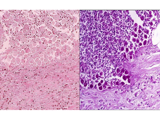
The left image shows several amebic trophozoites scattered among inflammatory cells in an ulcer bed. The right image shows a PAS stain, which stains glycogen in amebae deep red. Note that the trophozoites are distinctly larger than leukocytes (about 20-30 mm in diameter or several times larger than leukocytes). The organisms are distributed along the "cutting edge" of the ulcer.
Impression
Colon, amebic colitis and amebic trophozoites
Pathology Pointer
PAS stain is useful in detecting the presence of amebic organisms.
Preparation
Fixed; H&E stain; Fixed, stained with PAS stain
View
Light micrograph
Specimen
Colon
Image Credit
Katsumi M. Miyai, M.D., Ph.DDepartment of Pathology
School of Medicine
University of California, San Diego

