Features
Summary
Findings
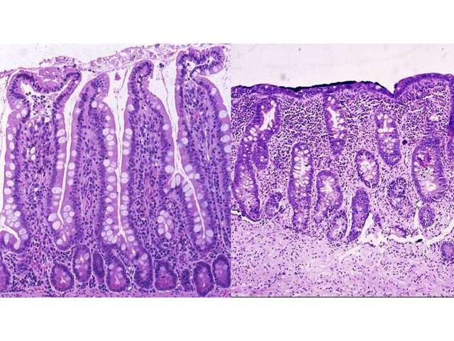
The left image is normal jejunum showing that the height of jejunal villi are 3-4 times the depth of the crypts. Compare this with the right image of Celiac Sprue. Here, the villi are totally lost due to severe atrophy, and crypts are irregular in their configuration. Marked chronic inflammation is seen in the lamina propria.
Impression
Normal jejunum; Jejunum, pathologic changes consistent with celiac sprue
Clinical Pathologic Correlation
Since this disease is caused by sensitivity to gluten, a component in wheat and related grains, removal of gluten from the diet leads to dramatic improvement. The disease also shows a strong genetic susceptibility, including familial clustering and association with the HLADQw2 histocompatibility antigen. There is a long-term risk of intestinal lymphoma, especially that of the T-cell type.
Pathology Pointer
Pathologic changes seen in celiac sprue are due to an immune reaction to gluten. Villous atrophy and chronic inflammation are not specific for this disease. Inflammatory cells are predominantly B lymphocytes and plasma cells. Malabsorption is thought to result from injury to the absorptive epithelium and from a decrease in absorptive surface area due to villous atrophy.
Preparation
Fixed, H & E stain
View
Light micrograph
Specimen
Jejunum
Image Credit
Katsumi M. Miyai, M.D., Ph.DDepartment of Pathology
School of Medicine
University of California, San Diego

