Features
Summary
Findings
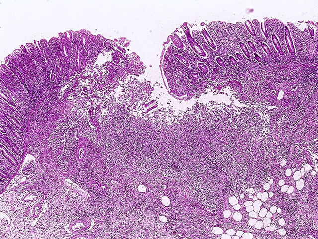
The ulcer shown in this image has a narrow opening through the mucosa and extends out laterally in the submucosa, undermining the mucosa and forming a "flask-shaped" defect.
Impression
Colon, amebic colitis
Preparation
Fixed, H & E stain
View
Light microscopy
Specimen
Colon
Image Credit
Katsumi M. Miyai, M.D., Ph.DDepartment of Pathology
School of Medicine
University of California, San Diego

