Features
Summary
Findings
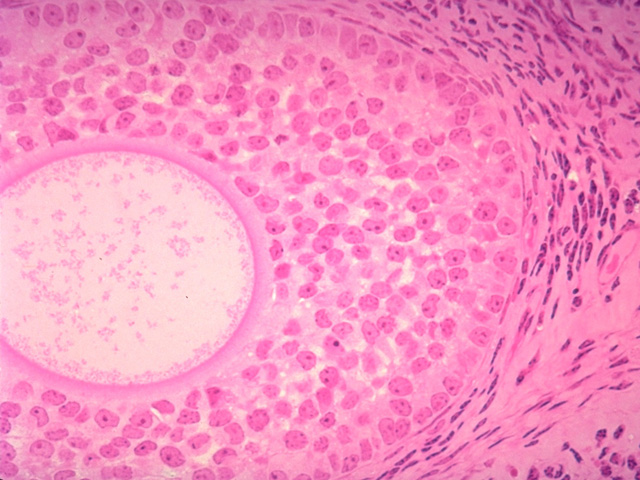
In this image of a late multilaminar primary follicle, the oocyte, and the surrounding zona pellucida and granulosa cells are clearly seen. Note the contrast between the granulosa cells and the surrounding ovarian stroma, consisting of theca interna and theca externa better seen in the next image.
Comment
The nucleus of the primary oocyte is not in the plane of section.
Preparation
Paraffin section, hematoxylin and eosin
View
High-power light microscopy
Specimen
Ovary
Image Credit
V. Eroschenko, Ph.D.Department of Biological Sciences
WAMI Medical Program
University of Idaho

