Features
Summary
Findings
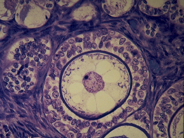
At very high power, the structure of a multilaminar primary follicle is revealed. Note the prominent oocyte containing a central nucleus with an eccentric nucleolus. A dense homogeneous zona pellucida surrounds the primary oocyte and separates it from the follicular cells, now called granulosa cells. A thin basement membrane separates the multilaminar primary follicle from the ovarian stroma.
Comment
The glycoproteinaceous zona pellucida contains cytoplasmic projections from both the primary oocyte and the inner layer of granulosa cells that are joined by gap junctions, facilitating transport of material to the developing oocyte. The clear areas in the cytoplasm of the oocyte are artifactual.
Preparation
Paraffin section; toluidine blue
View
High-power light microscopy
Specimen
Ovary
Image Credit
V. Eroschenko, Ph.D.Department of Biological Sciences
WAMI Medical Program
University of Idaho

