Features
Summary
Findings
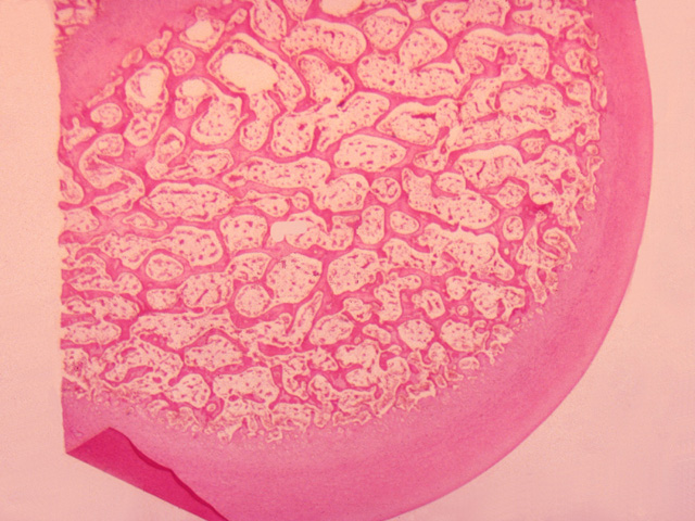
In this cross-section of decalcified bone, compact bone is seen surrounding spongy (cancellous) bone that consists of bone trabeculae and marrow.
Comment
Note the sieve-like arrangement of bone trabeculae and blood vessels scattered throughout the marrow.
Preparation
Paraffin section, hematoxylin and eosin
View
Low-power light microscopy
Specimen
Decalcified bone
Image Credit
Katsumi M. Miyai, M.D., Ph.DDepartment of Pathology
School of Medicine
University of California, San Diego

