Features
Summary
Findings
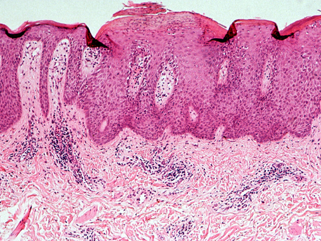
There is focal acanthosis up to several times the thickness of adjacent normal skin. The epidermal surface layer (stratum corneum) is parakeratotic (keratinized cells show residual nuclear staining). Dermal papillae extend close to the epidermal surface.
Impression
Psoriasis
Clinical Pathologic Correlation
Loss of the stratum granulosum (a water-proof layer)
results in a marked increase in the epidermal
permeability. Therefore, percutaneous administration of
drugs needs to be done with caution.
Pathology Pointer
Psoriatic acanthosis results from accelerated proliferation of basal layer cells, usually with loss of subsequent histologic differentiation (e.g. stratum granulosum often fails to develop). Tortuous and dilated blood vessels within extended dermal papillae give erythematous appearance to the lesion. Lesions bleed easily when the plaque is lifted (Auspitz sign).
Preparation
Fixed, H & E stain
View
Light micrograph
Specimen
Skin
Image Credit
Katsumi M. Miyai, M.D., Ph.DDepartment of Pathology
School of Medicine
University of California, San Diego

