Features
Summary
Findings
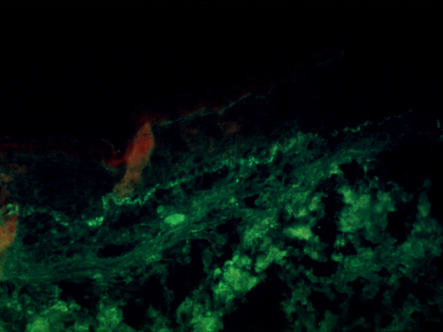
Note fluorescent deposits of C3 mainly along the dermalepidermal junction.
Impression
Complement C3 deposits, lupus erythematosus
Preparation
Immunohistochemical reaction for C3
View
Fluorescent light micrograph
Specimen
Skin
Image Credit
Terence O'Grady, M.D.Department of Medicine
School of Medicine
University of California, San Diego

