Features
Summary
Findings
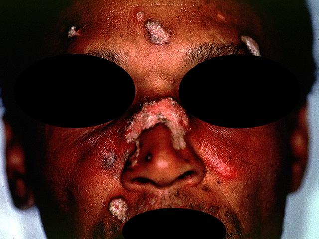
Another example of a patient with similar lesions as those seen in image #16 but with more severe involvement. Depigmentation is more distinct here.
Impression
Discoid lupus erythematosus
Preparation
Fresh
View
Clinical photograph
Specimen
Skin of the face
Image Credit
Terence O'Grady, M.D.Department of Medicine
School of Medicine
University of California, San Diego

