Summary
Findings
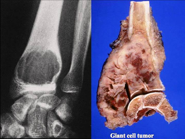
The radiograph (left) shows a mass in the distal radius. The right image is the gross appearance of a giant cell tumor. There is a bulging soft tissue mass that has destroyed the cortex. The predominance of foam cells can cause the white-yellow color.
Impression
Giant Cell Tumor
Clinical Pathologic Correlation
Giant cell tumors more commonly occur in the epiphysis and metaphysis of long bones, especially around the knee (distal femur and proximal tibia). It is relatively uncommon and usually benign thus treatment is with conservative surgery, e.g. curettage, however they can recur locally in up to 50% of cases. Age of incidence is 20-40yrs old.
Preparation
Fresh
View
Gross; radiograph
Specimen
Bone
Image Credit
Parviz Haghighi, M.D.Department of Pathology
School of Medicine
University of California, San Diego

