Features
Summary
Findings
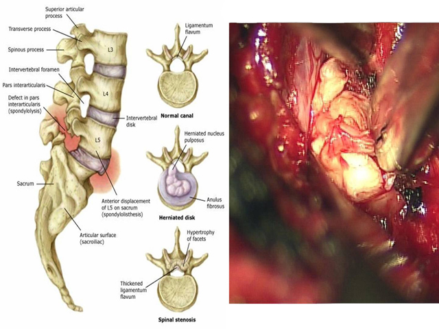
1) Diagram showing lateral and superior views of lumbar vertebra with normal anatomy, herniation of the nucleus pulposus, and spinal stenosis respectively; 2) Gross view of a herniated disk.
Impression
1) Diagram, Normal and Degenerated Intervertebral Disk; 2) Gross View of herniated disk
Clinical Pathologic Correlation
Intervertebral disks consist of fibrocartilage and are organized into an outer ring, the anulus fibrosis, and a central core, the nucleus pulposus. As the disk degenerates, i.e. dehydrates and loses resilience with age, the outer ring becomes more susceptible to tears, from either an acute event or gradually with repetitive strain. Large tears can permit the inner nucleus pulposus to herniate and compress nerve roots of the spinal cord.
View
Diagram; gross
Specimen
Herniated Disk
Image Credit
[Diagram]Richard A. Deyo, M.D., M.P.H. and James. N. Weinstein, D.O. Primary care: Low back pain. N Engl J Med. 2001 Feb 1;344 (5): 363-370. Copyright © [2001] Massachusetts Medical Society. All rights reserved.
[Gross]
Yu-Po Lee, M.D.
Department of Orthopedic Surgery
School of Medicine
University of California, San Diego

