Features
Summary
Findings
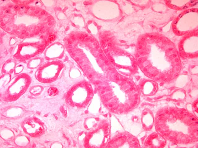
At higher power, the renal medulla shows collecting tubules and thick descending and ascending portions of the loops of Henle with simple cuboidal epithelium, and cross-sectional profiles of the thin loops of Henle with simple squamous epithelium. Scattered throughout the loose connective tissue of the renal interstitium are capillary profiles of the peritubular vascular network called the vasa recta.
Comment
Note that the epithelium of the collecting duct in the center of the image appears taller, almost columnar. This is due the tangential section through this duct.
Preparation
Paraffin section, hematoxylin and eosin
View
Medium-power light microscopy
Specimen
Renal medulla
Image Credit
V. Eroschenko, Ph.D.Department of Biological Sciences
WAMI Medical Program
University of Idaho

