Features
Summary
Findings
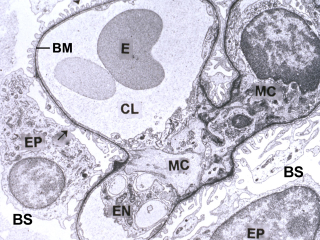
podocyte (EP)mesangial cell (MC)
capillary lumen (CL)
basal lamina (BM)
endothelial cell (EN)
erythrocyte (E)
Bowman's space (BS)
With transmission electron microscopy, the cellular detail of the renal corpuscle is further resolved. The glomerular capillary lumen containing erythrocytes is lined by an endothelium and covered by numerous processes (pedicels) of podocytes. Note the basal lamina between the endothelium and podocytes, and several mesangial cells within the glomerular matrix. Bowman's space is visible around the podocytes and their processes.
Comment
The filtration interface consists of a fenestrated endothelium, the interdigitating pedicels of the podocytes and the basal lamina produced by both. Mesangial cells are thought to maintain the functional integrity of the interface by phagocytotic removal of trapped material.
Preparation
Plastic section; uranyl acetate and lead citrate
View
Medium-power transmission electron microscopy
Specimen
Renal corpuscle
Image Credit
Katsumi M. Miyai, M.D., Ph.DDepartment of Pathology
School of Medicine
University of California, San Diego

