Summary
Findings
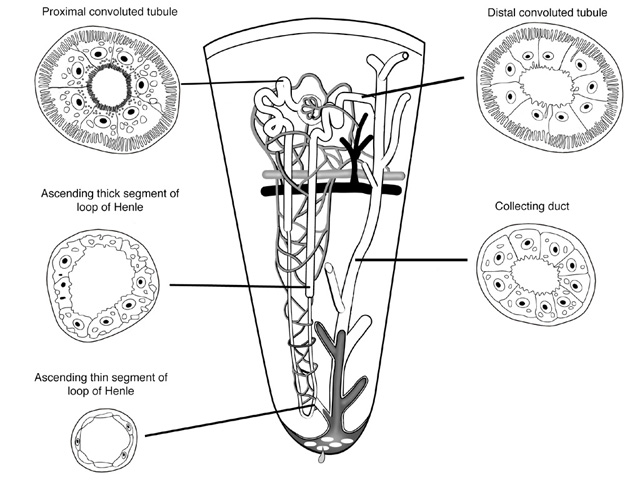
This diagram illustrates the important structural features of the nephron from the renal corpuscle to the distal convoluted tubule in the context of associated vasculature. Further, the connection of the nephron to the collecting duct system that coalesces into renal papillae is also shown. Note the regional differences in the epithelial lining and caliber of the renal tubule.
Comment
Blood carried to the glomeruli is filtered and the raw ultrafiltrate funneled to the proximal convoluted tubule where glucose, amino acids, small proteins, vitamins, sodium and water are reabsorbed. Tubular fluid leaves the cortex and enters the descending loop of Henle where water is passively removed and then the ascending loop of Henle where Na+ and Cl- are actively removed. Tubular fluid leaves the medulla and enters the distal convoluted tubule in the cortex where the salt and water balance between the urine and blood is adjusted with the help of the juxtaglomerular apparatus and renin-angiotensin-aldosterone system. Urine leaves the nephron and enters the system of collecting tubules and ducts that pass back through the medulla.
Specimen
Nephron
Image Credit
Andrew P. Mizisin, Ph.D.Department of Pathology
School of Medicine
University of California, San Diego
Katsumi M. Miyai, M.D., Ph.D
Department of Pathology
School of Medicine
University of California, San Diego

