Features
Summary
Findings
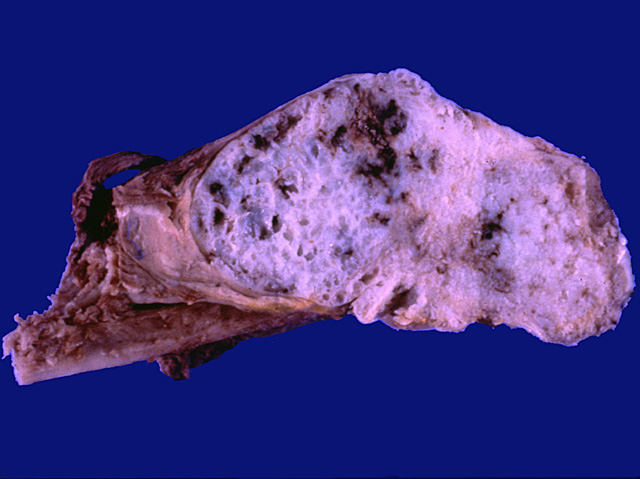
The cut section of a long bone, presumably the femur, reveals a tumor resembling cartilage (glistening and blue-grey in color) that has eroded through the cortex.
Impression
Chondrosarcoma
Clinical Pathologic Correlation
This tumor is second only to osteosarcoma in
frequency and occurs twice as often in males as in
females. It tends to occur in older people (peak
incidence in the 6th decade), and arises in central
portions of the skeleton, e.g. shoulder, pelvis,
proximal femur and ribs.
Pathology Pointer
Chondrosarcoma is a tumor composed of malignant mesenchymal cells that produce a cartilaginous matrix.
Preparation
Fresh
View
Gross specimen
Specimen
Lower extremity
Image Credit
Katsumi M. Miyai, M.D., Ph.DDepartment of Pathology
School of Medicine
University of California, San Diego

