Features
Summary
Findings
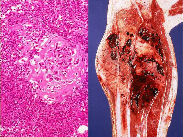
At high power, note the finely woven, eosinophilic osteoid surrounded by bone forming tumor cells with pleomorphic and hyperchromatic nuclei. On the right is a gross view of the tumor invading the long bone. Note a large, poorly-demarcated lesion situated within the metaphysis which has destroyed the bony cortex, extended inward into the bone marrow and outward into the adjacent soft tissue.
Impression
Osteosarcoma
Clinical Pathologic Correlation
This tumor usually occurs in young individuals (most often in the second decade) and arises most commonly around the knee (distal femur and proximal tibia). Diagnosis requires the identification of osteoid formation, which is the hallmark of this tumor.
Pathology Pointer
Osteosarcoma usually arises de novo, but it may occur as a complication of an underlying bone disorder.
Preparation
Fixed, decalcified, H & E stain; Fresh
View
Gross; Light Micrograph
Specimen
Neoplasm of the bone
Image Credit
Parviz Haghighi, M.D.Department of Pathology
School of Medicine
University of California, San Diego

