Features
Summary
Findings
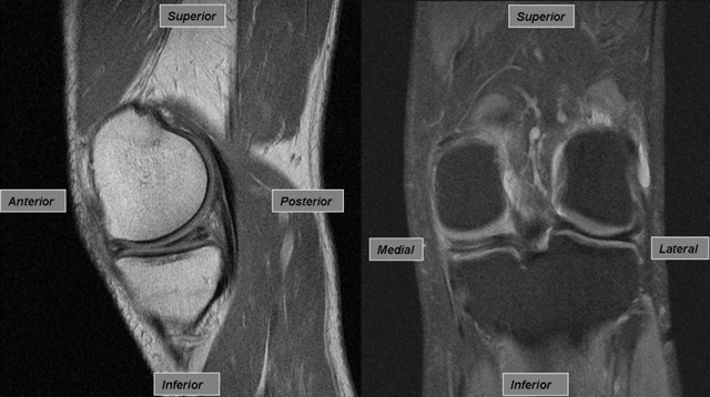
The left image is a sagittal view of a left knee with a medial meniscal tear. The tear appears as a white linear opacity between the articular surfaces of the distal femur and proximal tibia, where the menisci are located. This linear opacity extends to the articular cartilage of the tibia, thus supporting the diagnosis of a meniscal tear. Cartilage such as the meniscus appears black in this MRI. In a normal knee this region looks like a solid black triangle. The right image is an anterior-posterior view of the same left knee. The meniscal tear on the medial side appears opaque whereas the normal, lateral meniscus appears as a black line.
Impression
Meniscal Tear (medial) , Left Knee
Clinical Pathologic Correlation
The medial and lateral menisci are fibrocartilaginous pads that function to reduce stresses across the femoral condyles and tibial plateaus. A significant twisting injury to the knee can cause a traumatic tear, while degenerative tears in older patients may occur during simple activities. Traumatic tears can lead to knee swelling and stiffness in the first few days, and may be followed by mechanical symptoms such as locking, catching, or popping. Treatment typically consists of RICE (rest, ice, compression, elevation) along with pharmacotherapy for pain and physical therapy. However surgical management may be considered in certain cases (ex. young patients with significant tears, older patients with no response to conservative therapy).
View
MRI (magnetic resonance image)
Specimen
Left Knee
Image Credit
Douglas Chang, M.D., Ph.D.Department of Orthopedic Surgery
School of Medicine
University of California, San Diego

