Features
Summary
Findings
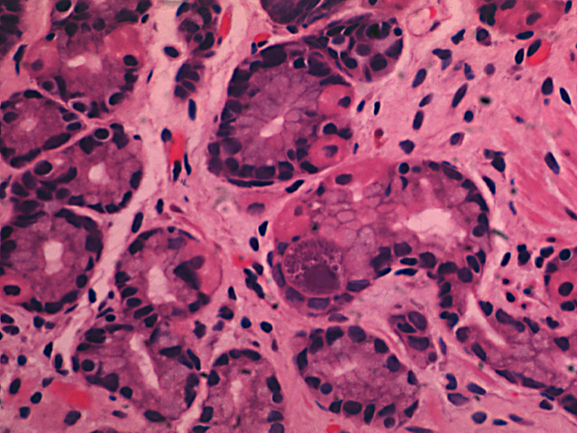
Note the large reddish-purple intranuclear inclusion in an enlarged gastric epithelial cell. Several smaller intracytoplasmic inclusions are also present.
Impression
CMV gastritis
Pathology Pointer
CMV inclusions are found in non-epithelial cells and, less commonly, in epithelial cells. An enlarged cell with a big, reddish-purple intranuclear inclusion surrounded by a clear halo and peripherally clumped chromatin is characteristic for CMV infection. Cells with similar but smaller intranuclear inclusions are also found in the herpes-varicella group of infections. Immunohistochemistry or DNA probes are required for a definitive diagnosis. The infected cells with inclusions are markedly enlarged, hence the term "cytomegalo".
Preparation
Fixed, H & E stain
View
Light microscopy
Specimen
Stomach
Image Credit
Katsumi M. Miyai, M.D., Ph.DDepartment of Pathology
School of Medicine
University of California, San Diego

