Features
Summary
Findings
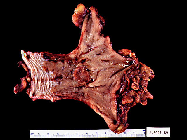
This image shows the distal esophagus and the proximal stomach. The white-grey segment of the esophagus is normal, whereas the light tan mucosa immediately proximal to the stomach is lined by metaplastic columnar epithelium called Barrett's esophagus. In the fresh, unfixed state, Barrett's esophagus appears salmon-pink in color. The round, disk-like lesion in the area of the metaplastic mucosa is a focus of adenocarcinoma.
Impression
Esophagus, Barrett's metaplasia with adenocarcinoma
Clinical Pathologic Correlation
Barrett's esophagus develops in about 10% of
individuals with gastroesophageal reflux. It is
associated with an 30-40 fold increased risk of
developing adenocarcinoma.
Pathology Pointer
Barrett's esophagus is defined as the replacement of normal stratified squamous epithelium by metaplastic columnar epithelium caused by long-standing gastroesophageal reflux.
Preparation
Fixed in formalin
View
Gross photograph
Specimen
Esophagus
Image Credit
Katsumi M. Miyai, M.D., Ph.DDepartment of Pathology
School of Medicine
University of California, San Diego

