Features
Summary
Findings
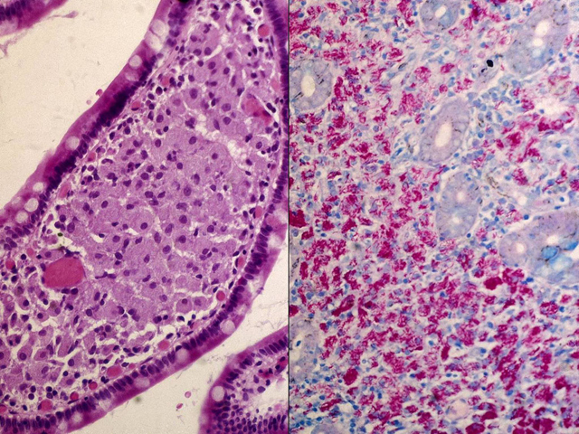
The high power view (left image) shows the lamina propria of a villus with numerous pale-pink "foamy macrophages". These macrophages are loaded with many red staining (acid-fast) bacilli (right image).
Impression
MAI Infection
Pathology Pointer
Foamy macrophages are seen in a variety of conditions. Therefore, more specific diagnostic studies other than H&E staining are required to identify the causative agent. A positive acid-fast stain is strongly suggestive for myobacterial infection. However, further microbiological study is needed for the definitive diagnosis of Mycobacterium avium intracellulare.
Preparation
Fixed, H & E stain; AFB stain
View
Light micrograph
Specimen
Small intestine
Image Credit
Katsumi M. Miyai, M.D., Ph.DDepartment of Pathology
School of Medicine
University of California, San Diego

