Features
Summary
Findings
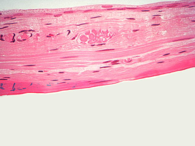
Again at high magnification, the bladder mucosa is shown but this time in an extremely distended state. Note the blood vessel in the lamina propria between the transitional epithelium and muscularis.
Comment
When the bladder is distended, cells of the transitional epithelium are extremely flattened and appear squamous.
Preparation
Paraffin section, hematoxylin and eosin
View
High-power light microscopy
Specimen
Urinary bladder
Image Credit
V. Eroschenko, Ph.D.Department of Biological Sciences
WAMI Medical Program
University of Idaho

