Features
Summary
Findings
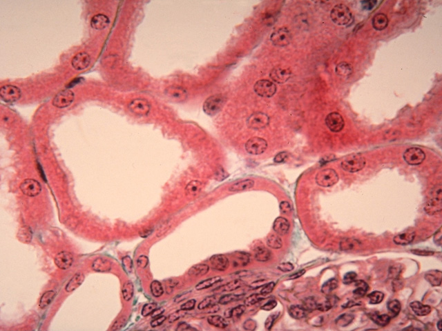
In this medium-power view, cross sections of renal tubules and a portion of a glomerulus with adjacent juxtaglomerular cells are seen. Two profiles of distal convoluted tubules are shown, one of which contains the closely spaced nuclei of the macula densa. Above the distal convoluted tubules are profiles of proximal convoluted tubules.
Comment
Proximal and distal convoluted tubules are distinguished by their relative number and luminal profile. Because the proximal convoluted tubule is longer than the distal convoluted tubule, more cross-sectional profiles are usually evident in the cortex. Further, because the brush border is better developed in the proximal convoluted tubule, the luminal profile appears irregular and "fuzzy", while that of the distal convoluted tubules is more clearly delineated.
Preparation
Paraffin section; Masson's trichrome
View
Medium-power light microscopy
Specimen
Renal tubules
Image Credit
V. Eroschenko, Ph.D.Department of Biological Sciences
WAMI Medical Program
University of Idaho

