Features
Summary
Findings
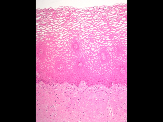
In this image of the mucosa of human vagina, the superficial layers of the stratified squamous nonkeratinized epithelium exhibit clear or empty cytoplasm. Because the vaginal wall was cut at an oblique angle, papillae from the underlying connective tissue appear as oval structures within the epithelium.
Comment
The empty appearance of cells in the superficial layers of the vaginal epithelium is due to increased glycogen accumulation, which is washed out during routine histological preparation. As epithelial cells are sloughed into the vaginal lumen, glycogen is metabolized into lactic acid by the vaginal bacterial flora, producing an acidic environment that helps restrict infection.
Preparation
Plastic section; hematoxylin and eosin
View
High-power light microscopy
Specimen
Vagina
Image Credit
V. Eroschenko, Ph.D.Department of Biological Sciences
WAMI Medical Program
University of Idaho

