Features
Summary
Findings
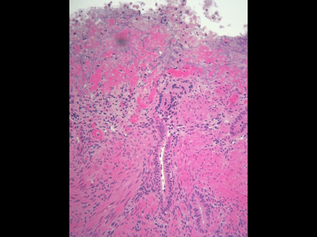
In this image of the menstrual uterus, remains of the functional layer are seen surrounded by cellular debris and pools of blood. The basal layer of the endometrium with portions of uterine glands remains intact.
Comment
If fertilization and implantation do not occur, the corpus luteum degenerates, and there is a rapid decline in estrogen and progesterone production. Loss of these hormones results in spasmodic constriction of spiral arteries that nourish the functional layer, causing ischemia and necrosis of this layer. When these arteries open again, the weakened vessel walls rupture and blood fills the surrounding connective tissue, causing the damaged functional layer to separate from the basal layer and be discharged in the uterine cavity as menstrual flow. Because the basal layer receives a separate blood supply, it is not damaged during menstruation. Cells and the remnants of uterine glands in this layer proliferate, resurface the endometrium and form a new functional layer during the proliferative phase.
Preparation
Paraffin section, hematoxylin and eosin
View
Medium-power light microscopy
Specimen
Uterus
Image Credit
V. Eroschenko, Ph.D.Department of Biological Sciences
WAMI Medical Program
University of Idaho

