Features
Summary
Findings
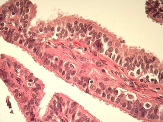
At very high-power, the cellular structure of a mucosal fold in the fallopian tube is seen. Note that the epithelium consists of ciliated and nonciliated (peg) cells. These cells rest on a lamina propria that forms the core of the folds and contains a nutritive capillary network.
Comment
In this section, the nonciliated cells are secretory, less numerous, appear squeezed between ciliated cells and provide nutritive products for the uterine tube. The ciliated cells are most numerous near the ovary and their cilia beat towards the uterus, facilitating movement of the ovum towards the uterus.
Preparation
Paraffin section; Masson's trichrome
View
High-power light microscopy
Specimen
Fallopian tube
Image Credit
V. Eroschenko, Ph.D.Department of Biological Sciences
WAMI Medical Program
University of Idaho

