Features
Summary
Findings
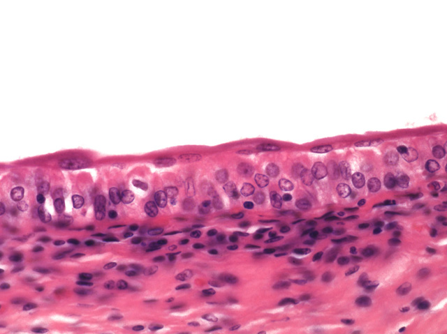
In contrast to slide #14, the apical cells of the transitional epithelium from this urinary bladder are thin and stretched, suggesting that this bladder was full when the tissue was fixed. The apical cells have mobilized the membrane plaques, allowing this stretched configuration.
Comment
Unlike the stratified squamous epithelium from thin skin, only the outermost cell layer in transitional epithelium is squamous in appearance and each cell contains a nucleus.
Preparation
Paraffin section, hematoxylin and eosin
View
Medium-power light microscopy
Specimen
Urinary bladder, distended
Image Credit
V. Eroschenko, Ph.D.Department of Biological Sciences
WAMI Medical Program
University of Idaho

