Features
Summary
Findings
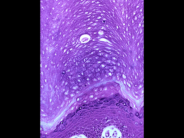
Shown here is a higher magnification of slide # 12. The area stained light purple is from the keratinized region of the epithelium where cells lack nuclei, while that stained dark purple is from the region with viable cells that contain nuclei. The light blue line that separates the two layers is actually a group of cells (stratum lucidum) with unique staining properties but no other significance.
Comment
The relative thickness of the keratinized layer gives a clue to the type of skin. Thick keratinized layers are found on the palms of the hands and soles of the feet. Thin keratinized layers are found on skin from the back, abdomen, arms, legs, etc.
Preparation
Paraffin section, hematoxylin and eosin
View
High-power light microscopy
Specimen
Thick skin
Image Credit
Katsumi M. Miyai, M.D., Ph.DDepartment of Pathology
School of Medicine
University of California, San Diego

