Features
Summary
Findings
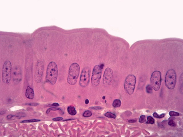
At much higher magnification, the brush border of the absorptive simple columnar epithelium is evident. Note that a portion of a large capillary filled with blood cells is seen in the subjacent lamina propria.
Comment
The brush border seen by light microscopy represents microvilli when viewed by electron microscopy. Microvilli greatly increase the apical absorptive surface area of the mucosal epithelium.
Preparation
Paraffin section, hematoxylin and eosin
View
High-power light microscopy
Specimen
Small intestine
Image Credit
V. Eroschenko, Ph.D.Department of Biological Sciences
WAMI Medical Program
University of Idaho

