Features
Summary
Findings
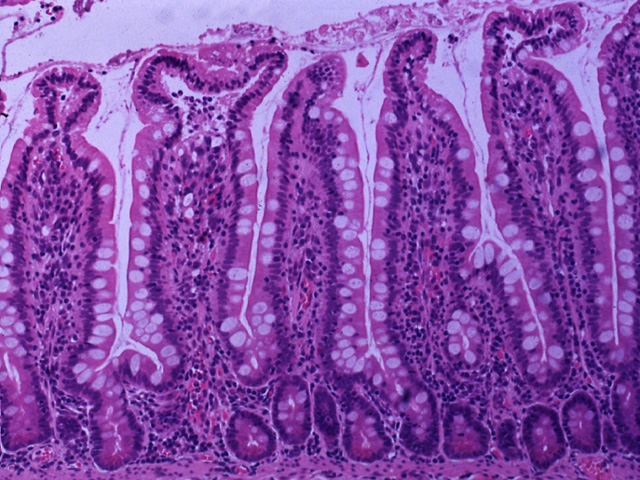
In this medium-power image, the relationship between intestinal glands and villi can be seen. The intestinal glands open into the intervillous spaces between the villi. Note that the characteristic simple columnar epithelium with numerous goblet cells lines the villi and extends into the intestinal glands. The loose connective tissue of the lamina propria forms the core of the villi and surrounds the intestinal glands. Located at the bottom of the intestinal glands are Paneth cells that are filled with numerous red granules.
Comment
Undifferentiated precursors of the absorptive epithelial cells, goblet cells, Paneth cells and enteroendocrine cells are found at the base of the intestinal glands.
Preparation
Paraffin section, hematoxylin and eosin
View
Medium-power light microscopy
Specimen
Small intestine
Image Credit
Katsumi M. Miyai, M.D., Ph.DDepartment of Pathology
School of Medicine
University of California, San Diego

