Features
Summary
Findings
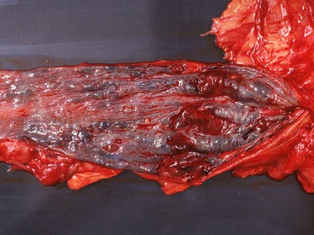
Note the markedly distended veins in the distal esophagus.
Impression
Esophageal varices
Clinical Pathologic Correlation
Esophageal varices are one of the major
complications of cirrhosis. With increased
mechanical resistance, portal venous blood is
diverted through collateral routes, e.g. esophageal,
hemorrhoidal, splenic, and umbilical veins. Massive
hemorrhage often results from esophageal varices
with serious consequences.
Preparation
Fresh
View
Gross photograph
Specimen
Esophagus
Image Credit
Katsumi M. Miyai, M.D., Ph.DDepartment of Pathology
School of Medicine
University of California, San Diego

