Features
Summary
Findings
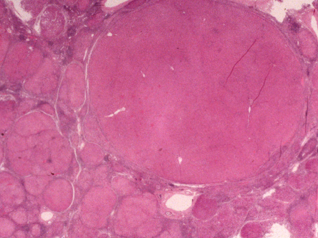
This image shows the key histologic and diagnostic features of cirrhosis, namely, fibrous septa, regenerative nodules, and extensive parenchymal involvement.
Impression
Hepatic cirrhosis
Clinical Pathologic Correlation
Clinical studies alone do not establish the
diagnosis of cirrhosis. Histopathologic
confirmation is required for a definitive diagnosis.
Pathology Pointer
Nodules shown in this image vary a great deal in size and some of the larger ones contain intact lobules. This type of cirrhosis, called macronodular cirrhosis, typically results from chronic hepatitis in which the distribution and severity of tissue injury are not uniform.
Preparation
Fixed, H & E stain
View
Light micrograph
Specimen
Liver
Image Credit
Katsumi M. Miyai, M.D., Ph.DDepartment of Pathology
School of Medicine
University of California, San Diego

